Introduction
The cardiovascular system is an intricate network of veins, arteries, and organs that work together to transport blood throughout the body. But what many people don’t realize is that the heart is the center of this system — it’s the organ responsible for pumping blood to the rest of the body. Understanding how blood travels through the heart is essential in order to comprehend the entire circulatory system.
In this article, we’ll explore the pathway of blood through the heart, from the right atrium and ventricle to the left atrium and ventricle and on to the pulmonary circulation and systemic circulation. We’ll also look at the differences between arteries and veins, how the valves work, and how various conditions can affect blood flow and cardiac output. Finally, we’ll provide a visual guide to help readers better understand how blood travels through the heart.
Step-by-Step Guide to the Path of Blood Through the Heart
The journey of blood through the heart begins with the right atrium, which is the upper chamber of the heart. From here, the blood passes through the tricuspid valve into the right ventricle. The right ventricle pumps the blood out of the heart and into the lungs, where it picks up oxygen. This is known as the pulmonary circulation.
Once the blood has been oxygenated in the lungs, it returns to the left atrium of the heart via the pulmonary veins. From the left atrium, the blood passes through the mitral valve into the left ventricle. The left ventricle then pumps the oxygenated blood out of the heart and into the systemic circulation — the network of arteries and veins that transport the blood to the rest of the body.
The blood then travels through the arteries to the capillaries, where the oxygen and nutrients are delivered to the cells. The deoxygenated blood is then collected by the veins and transported back to the heart, completing the cycle.

Exploring the Journey of Blood Through the Heart
To better understand the path of blood through the heart, it’s helpful to get an overview of the four chambers, the valves, and the differences between arteries and veins.
Overview of the Four Chambers
The heart is divided into four chambers: the right atrium, right ventricle, left atrium, and left ventricle. The right atrium receives deoxygenated blood from the body, while the left atrium receives oxygenated blood from the lungs. The right ventricle pumps the deoxygenated blood to the lungs, while the left ventricle pumps the oxygenated blood to the rest of the body.
How the Valves Work
The heart contains four valves that prevent the blood from flowing backward. The tricuspid valve is located between the right atrium and right ventricle, while the mitral valve is located between the left atrium and left ventricle. The pulmonic and aortic valves are located between the right and left ventricles, respectively, and regulate the flow of blood from the ventricles to the pulmonary artery and aorta.
Differences Between Arteries and Veins
Another important aspect of understanding the path of blood through the heart is knowing the differences between arteries and veins. Arteries carry oxygenated blood away from the heart and toward the body’s tissues, while veins carry deoxygenated blood back to the heart. The walls of arteries are thicker and more elastic than those of veins, which helps them withstand the higher pressure of the pumped blood.

An Overview of Blood Flow Through the Heart
Now that we’ve explored the four chambers of the heart and the roles of the valves, let’s take a look at the pathways of oxygenated and deoxygenated blood. Oxygenated blood is carried from the lungs to the left atrium via the pulmonary veins, then through the mitral valve into the left ventricle. From the left ventricle, the oxygenated blood is pumped out of the heart via the aortic valve and into the aorta, the main artery of the systemic circulation.
Deoxygenated blood enters the right atrium from the vena cava, the main vein of the systemic circulation. It then passes through the tricuspid valve into the right ventricle, which pumps the blood out of the heart and into the pulmonary artery. From there, the blood is transported to the lungs, where it is oxygenated and sent back to the left atrium.
It’s important to note that these pathways intersect at various points throughout the heart. For example, the aortic arch connects the aorta to the pulmonary artery, allowing some of the oxygenated blood to be diverted to the lungs. Similarly, the coronary sinus connects the vena cava to the right atrium, allowing some of the deoxygenated blood to mix with the oxygenated blood.
A Visual Guide to How Blood Travels Through the Heart
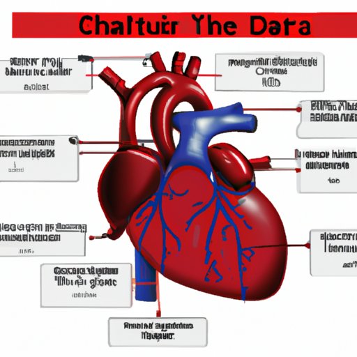
Visual aids are often helpful when trying to understand complex systems like the cardiovascular system. To that end, we’ve compiled a few diagrams and illustrations to help readers visualize the path of blood through the heart.
Diagrams of the Circulatory System
Diagrams of the circulatory system show the full pathway of blood from the heart to the lungs and back again. These diagrams include labels for the four chambers of the heart, the valves, and the major arteries and veins. They also show how the pathways of oxygenated and deoxygenated blood intersect at various points throughout the heart.
Illustrations of the Movement of Blood
Illustrations of the movement of blood through the heart are another useful visual aid. These illustrations show the flow of blood from the right atrium to the left ventricle, as well as the pathways of oxygenated and deoxygenated blood. They also indicate the direction of blood flow at each point in the pathway.

Understanding the Circulation of Blood Through the Heart
Now that we’ve looked at the path of blood through the heart, let’s explore how various conditions can affect blood flow and cardiac output. Cardiac output is the amount of blood that the heart pumps out in a minute, and it’s affected by factors such as heart rate and stroke volume. Heart rate is the number of times the heart beats per minute, while stroke volume is the amount of blood that is pumped out of the heart with each beat.
Various conditions can affect both heart rate and stroke volume. For example, exercise increases both heart rate and stroke volume, while certain medications can decrease heart rate and stroke volume. Additionally, certain health conditions, such as high blood pressure, can cause the heart to work harder, resulting in an increased cardiac output.
Finally, it’s important to note that the heart plays a crucial role in regulating blood pressure. As the heart pumps blood, it creates pressure on the walls of the arteries. This pressure is known as systolic blood pressure, and it decreases as the blood moves away from the heart. Diastolic blood pressure is the pressure in the arteries when the heart is resting between beats.
Conclusion
In this article, we explored the path of blood through the heart and the impact of various conditions on blood flow and cardiac output. We started by looking at the four chambers of the heart, the valves, and the differences between arteries and veins. Then, we provided an overview of the pathways of oxygenated and deoxygenated blood, followed by a visual guide to help readers better understand the circulation of blood through the heart.
To summarize, the journey of blood through the heart begins with the right atrium, where it is received from the vena cava. From here, the blood passes through the tricuspid valve into the right ventricle, which pumps the blood out of the heart and into the lungs. In the lungs, the blood is oxygenated and transported back to the left atrium via the pulmonary veins. From the left atrium, the blood passes through the mitral valve into the left ventricle, which pumps the oxygenated blood out of the heart and into the systemic circulation.
Various conditions can affect the flow of blood through the heart and the cardiac output. Exercise increases both heart rate and stroke volume, while certain medications can decrease heart rate and stroke volume. Additionally, certain health conditions, such as high blood pressure, can cause the heart to work harder, resulting in an increased cardiac output.
By understanding how blood travels through the heart, we can gain a better appreciation for the intricacies of the cardiovascular system and the importance of keeping our hearts healthy.
(Note: Is this article not meeting your expectations? Do you have knowledge or insights to share? Unlock new opportunities and expand your reach by joining our authors team. Click Registration to join us and share your expertise with our readers.)
